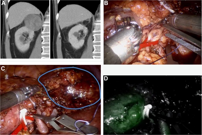
Figure 1.
Selective clamping of three arterial branches supplying the dorsal right-sided 6 cm upper pole RCC.
Notes: (A) Contrast-enhanced CT image demonstrating the upper pole 6 cm tumor of the right kidney preoperative and 6 months postoperative. (B) A dorsal segmental branch is clamped. The renal artery is put on tension with a vessel loop to improve exposure of the anterior upper pole arterial branches and a second bulldog is prepared to clamp. (C) Selective clamping of another two arterial branches supplying the dorsal right-sided 6 cm upper pole RCC. The lower pole is visible on the left side of the image. Gerotas fascia is covering the mid portion of the kidney. The tumor is encircled on the right side. (D) After application of indocyanine green, the perfusion of the lower pole of the kidney is visible, whereas the upper pole tumor is not perfused and therefore not visible.
Abbreviations: RCC, renal cell carcinoma; CT, computed tomography.

