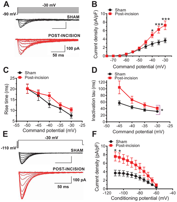
Fig. 1. Effects of plantar skin incision on T-current kinetics and density in rat DRG cells.
A. Families of T-currents evoked in representative DRG cells from sham and incised rats were used to generate current-voltage (I-V) relationships. B. Average peak T-current densities calculated from I-V curves were increased more than two-fold in the post-incision group (n=20 cells, 10 animals) compared with the sham group (n=14 cells, 10 animals) (***P<0.001, *P<0.05 two-way RM-ANOVA followed by Sidak’s post-hoc test; treatment: F(1,32)=5.52, p=0.02; interaction: F(9,288)=6.72, post-hoc: p=0.017 and p<0.001). C. Time-dependent activation (10-90 % rise time) measured from I-V curves in DRG cells was not significantly affected by plantar skin incision (n=14 cells from 10 sham animals; n=20 cels from 10 animals post-incision; two-way RM-ANOVA; p=0.3). D. Inactivation time constant measured from I-V curves was significantly slower approximately two-fold after plantar skin incision (n=14 cells, 10 sham animals; n=20 cells, 10 animals post-incision; *P<0.05 two-way RM-ANOVA, treatment: F(1,32)=4.33, p=0.04; interaction: F(3, 96)=2.36, p=0.08). E. Families of T-currents evoked in representative DRG cells by steady-state inactivation protocols in sham (back traces) and incised (red traces) groups. F. T-current densities from steady-state inactivation curves post-incision (n=21 cells, 10 animals) were significantly increased when compared to sham (n=14 cells, 10 animals) (two-way RM-ANOVA followed by Sidak’s post-hoc test; interaction: F(10, 330)=6.78, p<0.0001; post hoc: p=0.026, p=0.041; treatment: F(1,33)=4.15, p=0.049).

