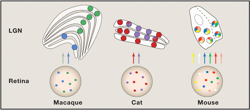
Figure 1. Organization of Visual Streams in Different Species.

Colored circles represent different retinal ganglion cell (RGC) types and their target relay neurons in the lateral geniculate nucleus (LGN). Mixing of stream s rarely occurs in m acaque and, to a limited extent, in cats. However, the results of the current study suggest that many more RGCs converge on individual relay cells in mouse LGN. Whether these RGCs are different functional subtypes remains unknown. Structures are not drawn to scale.
