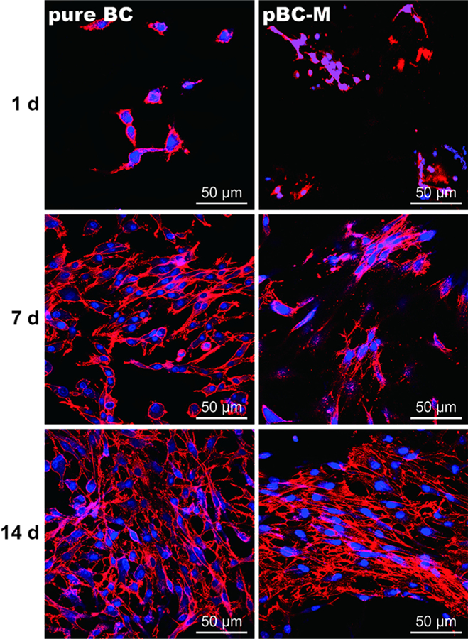
Fig. 6.

Fluorescence micrographs of chondrocytes cultivated on pure BC and pBC-M scaffolds after 1, 7 and 14 days. Actin filaments were labeled with rhodamine phalloidin and detected at an excitation wavelength of 632 nm and an emission wavelength 590 nm using epifluorescence microscopy. Cell nuclei were labeled with DAPI (blue) and detected at an excitation wavelength of 488 nm and an emission wavelength of 515 nm using epifluorescence microscopy.
