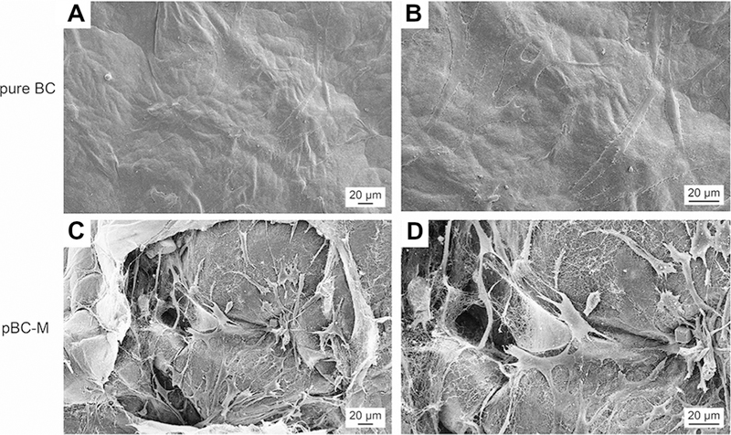
Fig. 7.

Chondrocyte cell adhesion on BC and pBC-M scaffolds after cultivation for 14 days. (A) Chondrocytes attached to the surface of pure BC scaffold. (B) Higher magnification images based on image in (A). (C) Cells spread around or in the pores of pBC-M scaffolds after seeding. (D) A higher magnification image of a region in (C).
