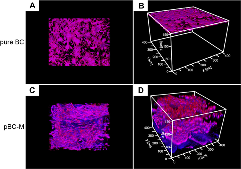
Fig. 8.

Distribution of chondrocyte cells seeded on BC and pBC-M scaffolds after 14 days. (A) Top-down view and (B) oblique view of fluorescence micrographs of chondrocytes seeded on BC and incubated for 14 days. (C) Top-down view and (D) oblique view of cells seeded on pBC-M substrates and cultivated for 14 days. Images were acquired on a laser scanning confocal microscopy and analyzed using ImageJ.
