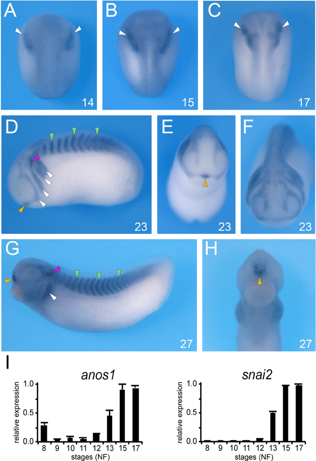
Figure 1: Developmental expression of anos1 by whole-mount ISH.

(A-C) At the neurula stage (NF stage 14–17), anos1 is detected in the prospective neural crest territory (white arrowheads). (D-F) At stage 23, anos1 is now more broadly expressed, to include the somites (green arrowheads), otic vesicle (red arrowhead), the anterior pituitary (yellow arrowhead) in addition to the branchial arches (white arrowheads). (G-H) Later in development (NF stage 27) anos1 persists in all these tissues. (A-C) dorsal views, anterior to top. (D, G) lateral views, dorsal to top, anterior to left. (E, F, H) frontal views, dorsal to top. The embryonic stages (NF) are indicated in the lower right corner of each panel. (I) Relative expression levels of anos1 and snail2 analyzed by qRT-PCR at the indicated stages. The values were normalized to odc1.
