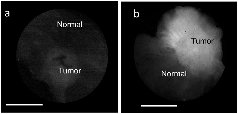
Figure 1:
LUM Imaging System images of transected tumors with and without LUM015 injection: (a) Transected lumpectomy specimen from Patient 5, IDC and DCIS, no LUM015 with tumor:normal signal ratio of 1.9 (b) Transected tumor specimen from Patient 8, IDC and DCIS, 0.5mg/kg LUM015 with tumor:normal signal ratio of 4.8. Images are plotted on linear brightness scales for which the minimum pixel value is black and the maximum pixel value is white. Scale bars = 1 cm.

