Figure 2:
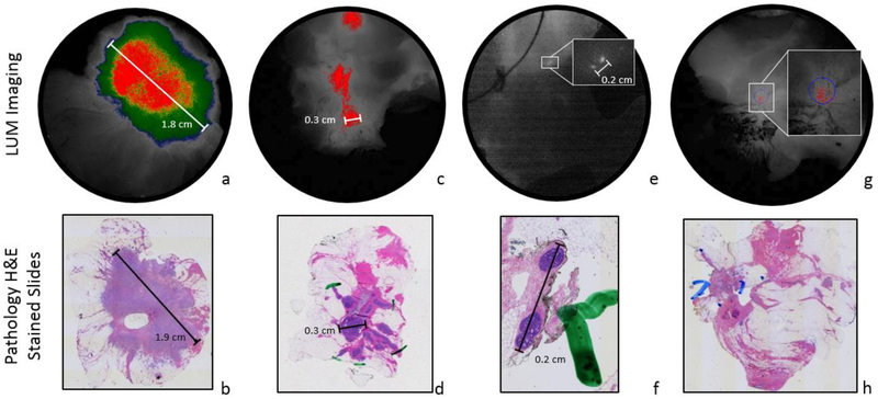
(a-b) Fluorescent image captured from transected ex vivo resected IDC mass from Patient 8. Pathology slide taken from same resected mass; the oval hole within the mass is a processing artifact
(c-d) Fluorescent image captured from transected ex vivo resected DCIS specimen in Patient 7. Pathology highlights evidence of 3 mm area of DCIS
(e-f) Ex vivo imaging of medial margin from patient 7. Two small fluorescent features appear, enlarged in the inset. Two foci of DCIS appear in the corresponding pathology slide.
(g-h) Lumpectomy transection LUM Image and corresponding H&E stained slide from patient 6. Pathology report defined lesion as invasive mammary carcinoma with mixed ductal and lobular features with DCIS within the invasive carcinoma.

