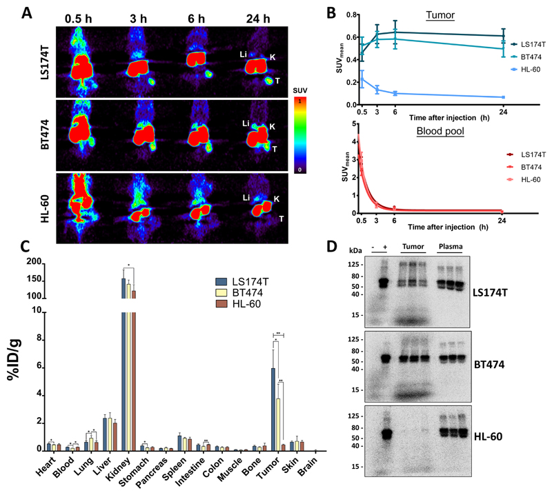
Fig. 3.
Uptake of 89Zr-AMG211 in LS174T (n = 6), BT474 (n = 6) or HL-60 (n = 6) tumor bearing mice. A) Representative coronal small-animal PET images up to 24 hours after injection of 10 µg 89Zr-AMG211. Li = liver; K = kidney; T = tumor. B) Quantification of tumors (upper panel) and blood pool (lower panel). Ex vivo C) biodistribution and D) SDS-PAGE autoradiography 89Zr-AMG211 24 hours after injection. + : 89Zr-AMG211 prior to injection; - : free 89Zr only; tumor: lysates of 3 different tumor bearing mice; plasma: plasma samples from corresponding mice. Data are mean ± SD. * P ≤ 0.05; ** P ≤ 0.01.

