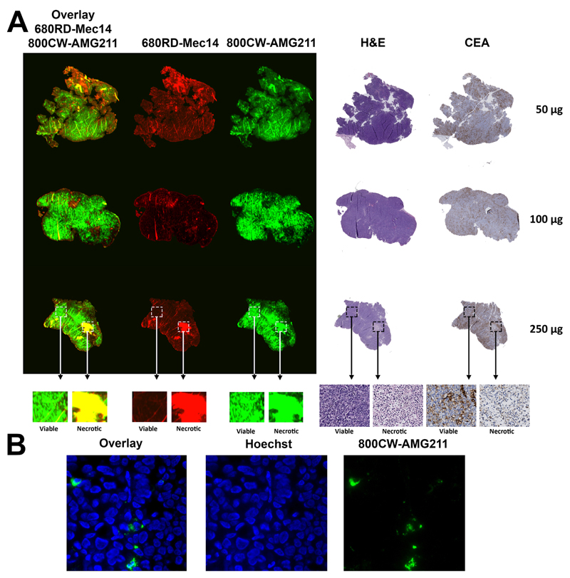
Fig. 5.
Intratumoral distribution of escalating doses of co-injected 800CW-AMG211 and 680RD-Mec14 (50, 100 or 250 μg) in LS174T tumors. (A) Macroscopic fluorescent imaging of 800CW-AMG211 (green) and 680RD-Mec14 (red) distribution, with overlapping signal (yellow) in necrotic tissue as visualized by hematoxylin and eosin (H&E). 800CW-AMG211 mainly localizes to viable tissue according to H&E with concordant CEA immunohistochemical staining. (B) Fluorescence microscopy images (630×), visualizing membrane and/or cytoplasmic localization of 800CW-AMG211 (green) and Hoechst stained nuclei (blue).

