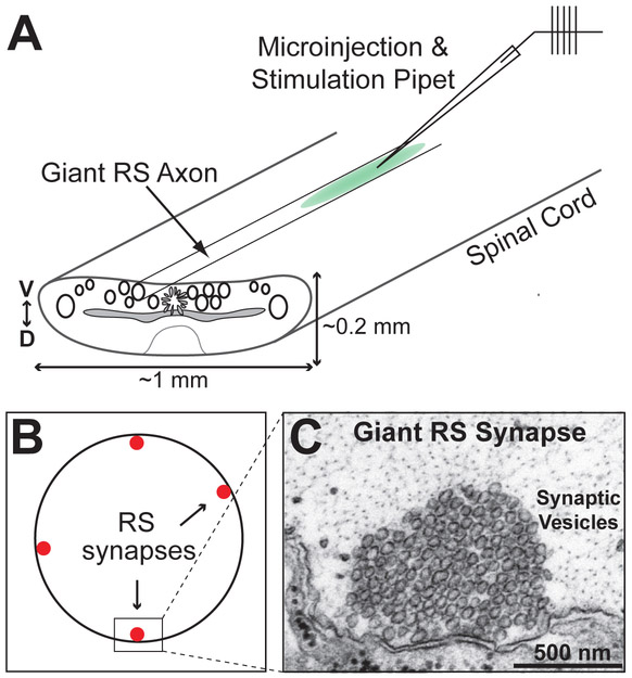
Figure 3.
Acute perturbations of CME at lamprey giant RS synapses. A. Diagram of the lamprey spinal cord and axonal microinjection. D=dorsal; V=ventral. B. Cross-section of a giant RS axon showing the synapses along the perimeter. C. An electron micrograph of an unstimulated lamprey giant RS synapse showing a large pool of tightly-clustered synaptic vesicles.

