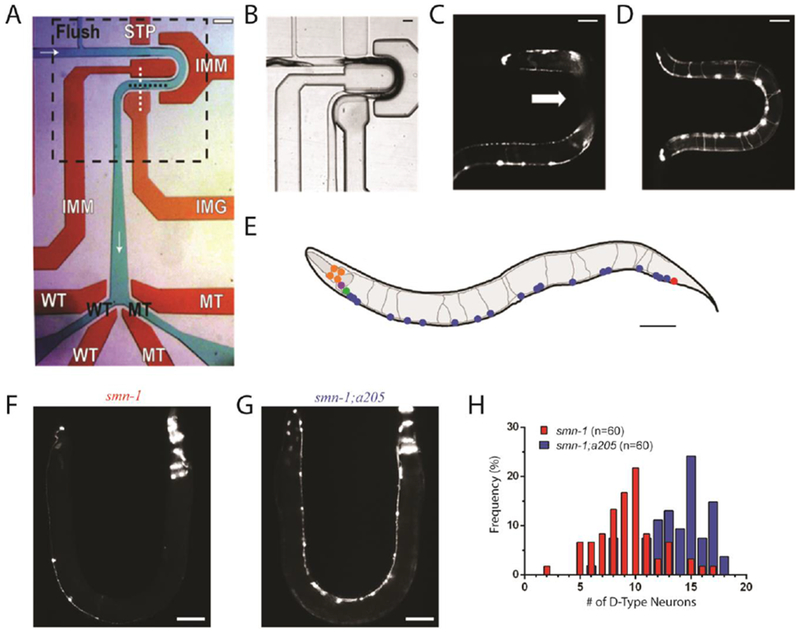
Fig. 1.

Single layer microfluidic device. (A) Device used for on-chip characterization and automated sorting of C. elegans. Flow layer is shown in green with black text, valve control layer shown in red with white text. Fluid flows from left to right, top to bottom, and is marked by white arrows. Flush channel, wild-type (WT), and mutant (MT) channels are all labelled. STP is the stop valve, IMM are the immobilization valves, IMG is the imaging valve, and WT and MT are the wild-type and mutant valves. Imaging area shown with dashed black box. Scale bar is 200μm. (B) Imaging area shown in panel A demonstrating valve expansion into flow layer and use of two immobilization valves to limit animal movement. Actuating the valve control layer simultaneously routes fluid flow and obstructs animal passage during device operations such as loading (shown) and imaging. (C) Example image of an animal body obstructed by immobilization valves. Device used for this image does not have a height difference in the imaging area. White arrow indicates area obscured from camera. (D) Example image of an animal body not obstructed by immobilization valves. Device used for this image does include a height difference of 20 μm in the imaging area. Scale bar for panels B-D is 70μm. (E) A rendering of a representative image of an adult worm carrying gbIs4[Punc-25::smn-1(RNAi sas); Pchs-2::GFP] [31] and oxIs12[Punc-47::GFP] transgenes, whose expression is visible in panels C, D, F and G. D-type motor neurons are shown in blue, cells expressing Pchs-2::GFP co-injection marker in orange, other GABA neurons where smn-1 is not silenced in red, green and purple. Image not to scale. Scale bar is 70μm (F) Image of one of the most severe example of smn-1(RNAi sas) transgenic animals with 2 out of 19 visible D-type motor neurons. (G) Image of allele smn-1;a205 with 16 out of 19 visible D-type motor neurons. Scale bar for panels F and G is 70μm. (H) Histogram demonstrating differences between motor neuron distributions within the two populations.
