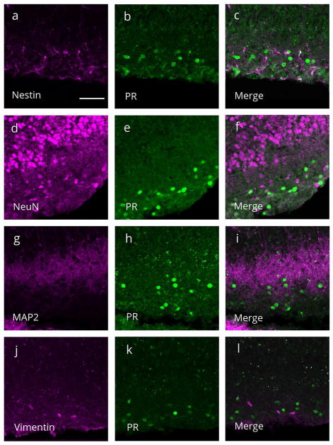
Figure 4.
Representative confocal images of single and merged channels through the molecular layer of the dentate gyrus on postnatal day 7 stained for progesterone receptor (PR) immunoreactivity (green) and (a–c) nestin-ir (magenta), a marker for neural precursor cells, (d–f) NeuN-ir (magenta), a marker for mature neurons, (g-i) MAP2-ir (magenta), a neuronal marker, or (j–l) vimentin-ir (magenta), a radial glial cell marker. (c,f,i,l) PR-ir was not colocalized with any of the markers. Scale bar: 50Om.

