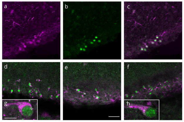
Figure 5.
Virtually all progesterone receptor (PR) cells expressed caltetinin and reelin. Representative confocal images of single and merged channels through the molecular layer of the dentate gyrus stained for (a) calretinin (magenta) (b) PR (green) and (c) colocalization of PR-ir and calretinin-ir. (d–f) colocalization of PR-ir (green) and reelin-ir (magenta). (g.h) PR/reelin expressing neurons had a ‘tadpole’ morphology typical of Cajal-Retzius neurons. (a-f) Scale bar: 50Om. (g,h) Scale bar: 10Om.

