
Figure 3.
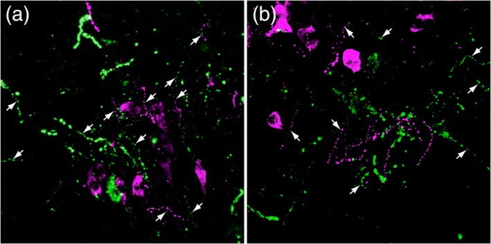
Epifluorescent dual localization of GLP‐1+ (green) and PrRP+ (magenta) neurons and processes in two different fields (a, b) within the rat caudal nucleus of the solitary tract, demonstrating the overlapping distribution of these separate neural populations. Both neural populations give rise to thick dendritic processes and thin varicose axons (arrows) [Color figure can be viewed at http://wileyonlinelibrary.com]