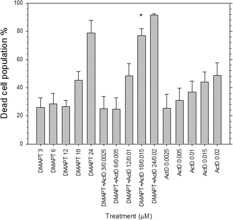
Figure 3.
Live-Dead FDA-PI assay in Panc-1 cells 48h post drug treatment was determined by flow cytometry. Treatment concentrations were chosen based on the respective GI50 values of DMAPT and ActD, and maintaining a constant ratio for the drug combination concentrations. Live and dead cell populations were detected by incubating Panc-1 cells with FDA at 37°C and PI at room temperature, respectively, in CO2-independent media.
(*p<0.05 vs each individual drug)

