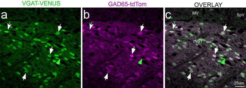
Figure 1.
Confocal micrographs (single Z level) of an rNST section from a mouse that was a cross between a GAD65-tdTom VGAT-Venus mouse. This section was approximately half-way between the rostral pole of the nucleus and the level at which the NST merges with the IVth ventricle. The photo centered on the regions of the rostral central and ventral subdivisions. White arrows illustrate examples of doubly-labeled neurons that were approximately equally bright for Venus and td-Tom; the green arrow denotes a singly-labeled VGAT neuron. The arrow with the white head and green tail points to a doubly-labeled neuron with more intense Venus than td-Tom label; examples of the converse were also observed, though none is obvious at this Z level. Scale = 25µm. (Abbreviations- MV: medial vestibular nucleus, rNST: rostral NST, SpV: spinal vestibular nucleus.

