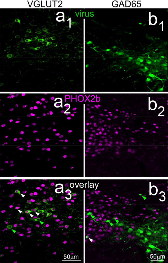
Figure 7.
Confocal photomicrographs (maximum intensity projections, two z levels, z=1µm intervals) near the centers of cre-dependent viral injection sites in VGLUT2 (a1 – a3) and GAD65 (b1 – b3) therelationship with PHOX2b staining. As described in the text, few virally-labeled neurons in the GAD65-cre mice were double-labeled (right overlay: b3), whereas a majority of those in the VGLUT case were double-labeled (left overlay: a3). Arrowheads show examples of neurons singly-labeled with virus (green) or double-labeled with PHOX2b and virus (white).

