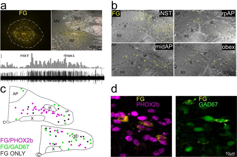
Figure 9.
Example of retrograde tracing in a GAD67-EGFP+ mouse (case ISM4) that received an injection of Fluorogold (FG), near the rostral pole of NST. Fluorogold-labeled neurons are pseudocolored yellow in all panels. a. Top panels: Photomicrographs of the injection site. Outlines on the left panel delineate the core (inner outline) and less densely-labeled shell (outer outline) of the injection site; the corresponding measurement appears in figure 10. The right panel shows the injection site superimposed upon a darkfield image of the nucleus. The core was mostly confined to NST, but some labeled cells in the shell extended into the vestibular nucleus dorsally and the reticular formation ventrally. Lower panel: Peristimulus-time histogram and raw neural record depicting the multiunit response to a taste mixture applied to the anterior tongue and recorded through the injection pipette. Scale bar for histogram: 5 spikes/500ms. b. Maximum intensity projections (2µm intervals) of confocal photomicrographs of retrograde labeling (yellow) superimposed upon corresponding DIC images extending from iNST to a level caudal to obex. c. Plots of retrogradely labeled neurons from a mid-level of the AP and obex (from a different series than in a and b) that was immunostained for PHOX2b. Retrogradely-labeled neurons are color-coded for PHOX2b (pink), GAD67 (green) or retrogradely-labeled only (grey). No neurons, including those retrogradely labeled, were double-labeled for PHOX2b and GAD67. d. Higher-magnification photomicrographs of retrogradely-labeled neurons double-labeled for GAD67 or PHOX2b. The arrowheads indicate the same neurons denoted in c. Abbreviations- AP: area postrema, C: Central canal, FG: Fluorogold, M: medial vestibular nucleus, RF: reticular formation, SpV: spinal vestibular nucleus, st: solitary tract, X: dorsal motor nucleus of the vagus, XII: hypoglossal nucleus.

