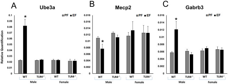
Fig. 3. WT male FAE litters showed autistic gene expression changes in the frontal cortex.
Relative quantification measured by real time RT-PCR indicates an increase in Ube3a (A), Gabrb3 (C) and decrease in Mecp2 (B) transcript levels in the frontal cortex of WT adult male litters of EtOH consuming mother. Ube3a (A), Gabrb3 (C) and Mecp2 (B) transcript levels were unaffected in TLR4−/− litters of EtOH consuming mothers. Values are mean ± SEM (n = 4 litters). Asterisk indicates the value that is significantly (p <0.05) different from corresponding control value. WT and TLR4−/− female FAE litters showed no effect on autistic gene expression in the frontal cortex. Relative quantification measured by real time RT-PCR indicates no effect on the mRNA expression levels of Ube3a, Mecp2 and Gabrb3 in the frontal cortex of WT and TLR4−/− adult female litters of EtOH consuming mother.

