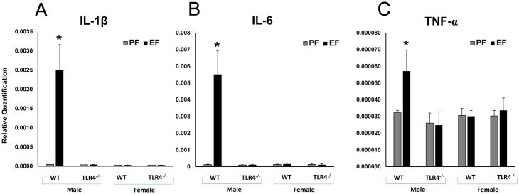
Fig. 4. WT male FAE litters showed increased proinflammatory cytokine gene expression in the frontal cortex.
Relative quantification measured by real time RT-PCR indicates an increase in IL-1β (A), IL-6 (B) and TNF-α (C) transcript levels in the frontal cortex of WT adult male litters of EtOH consuming mother. IL-1β (A), IL-6 (B) and TNF-α (C) transcript levels were unaffected in TLR4−/− litters of EtOH consuming mothers. Values are mean ± SEM (n = 4 litters). Asterisk indicates the value that is significantly (p <0.05) different from corresponding control value. WT and TLR4−/− female FAE litters showed no effect on inflammatory cytokine gene expression in the frontal cortex. IL-1β, IL-6 and TNF-α transcript levels were unaffected in WT and TLR4−/− litters of EtOH consuming mothers.

