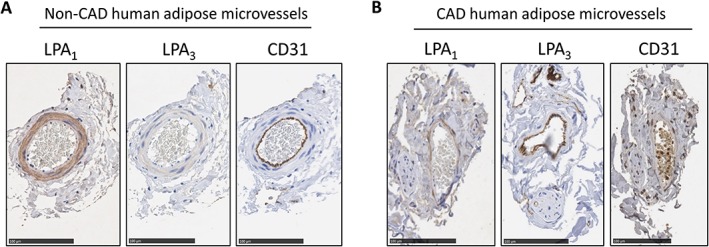
Figure 4.

LPA1 and LPA3 receptor protein expression in intact non‐CAD and CAD arterioles. (A) Immunohistochemical analyses for LPA1 and LPA3 receptors from vessels isolated from an individual without CAD. GAPDH used for loading control. (B) Immunohistochemistry analyses for LPA1 and LPA3 receptors in vessels from an individual with CAD. CD31 staining used to identify endothelial cell layer. Specificity of the antibody was determined by removal of the primary antibody (not shown). Bar for all images = 100 μm.
