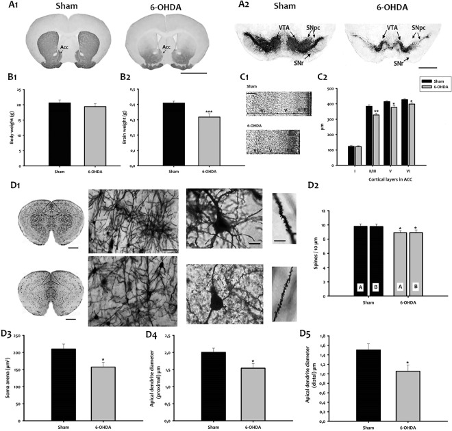
Figure 1.
Morphological characteristics of 6-OHDA mice. TH immunohistochemistry in the striatum (A1), and ventral midbrain (A2) of adult mice. Bar = 1000 μm. (B1,B2) Body and brain weight in grams. (C1). Nissl staining of the ACC. Bar = 400 μm. (C2). Thickness of cortical layers from I to VI (μm). Values are represented as mean ± SEM (*p < 0.05; **p < 0.01; ***p < 0.001; t-test; n = 7). (D1) (Left) Representative micrographs of mouse brains stained with Golgi Cox. Bar = 1000 μm. (Center left) Higher magnification images of Golgi-Cox stained brain sections from sham (top) and 6-OHDA (down) mice are shown. Bar = 100 μm. (Center right) Golgi staining of layer II-III pyramidal neurons (ACd) in sham (top) and 6-OHDA mice (down). Bar = 15 μm. (Right) Micrographs of Golgi-stained neurons showing dendrites of sham (top) and 6-OHDA (down). Bar = 25 μm. (D2) Dendritic spine density of apical and basal dendrites in the ACd neurons of the investigated experimental groups. A/B, apical/basal dendrites. (D3) Area of neuronal cell bodies. D4 Apical dendrite diameter (proximal) at 10 μm from the soma. (D5) Apical dendrite diameter (distal) at 100 μm from the soma. Bars represent mean ± SEM (*p < 0.05). Acc, accumbens nucleus; ACd, dorsal anterior cingulate cortex; SN, substantia nigra; SNpc, SN pars compacta; SNr, SN pars reticulata; VTA, ventral tegmental area.

