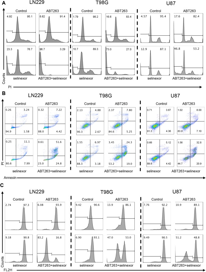
Figure 2.
The combination treatment of ABT263 and selinexor shows features of apoptotic cell death. (A) LN229, T98G and U87 GBM cells were treated with ABT263, selinexor or the combination. After 72 h, cells were harvested, fixed, stained with propidium iodide and analyzed by flow cytometric analysis for DNA – fragmentation. Shown are representative flow cytometry plots. (B) The same set of cell lines as in A were treated with the indicated drugs and the same conditions (except for the incubation time, which was 24 h). Thereafter, cells were stained with Annexin V/Propidium iodide and analyzed by multi-parametric flow cytometry. Shown are representative plots. (C) The same set of cell lines as in A were treated with the indicated drugs and the same conditions (except for the incubation time, which was 24 h). Thereafter, cells were stained with TMRE and analyzed by flow cytometry. Shown are representative flow plots.

