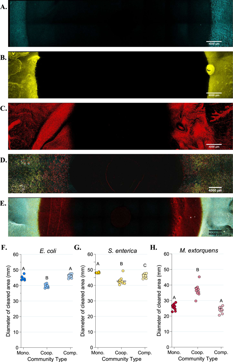
Fig. 5.
Fluorescent microscopy images of Petri plates with ampicillin antibiotic discs. An AZ100 confocal fluorescent macroscope at ×3.40 magnification was used to image 12 × 2 fields of view of each Petri plate to visualize E. coli (CFP, in blue), S. enterica (YFP, in yellow) or M. extorquens (RFP, red). a–e Representative images of E. coli monoculture (a), S. enterica monoculture (b), M. extorquens monoculture (c), cooperative community (d), and competitive community (e). Quantification of the diameter of the species-specific zone of clearing for E. coli (f), S. enterica (g), and M. extorquens (h) in each growth condition was performed in Elements software. The average of three technical replicate diameters was calculated to obtain a single measurement. At least 6 biological replicates were obtained for each species/growth condition. Pairwise comparisons of median diameter of clearing were performed using a Mann–Whitney U test, with a Bonferroni correction applied for three multiple comparisons. Significant differences are noted by different letters above each cluster

