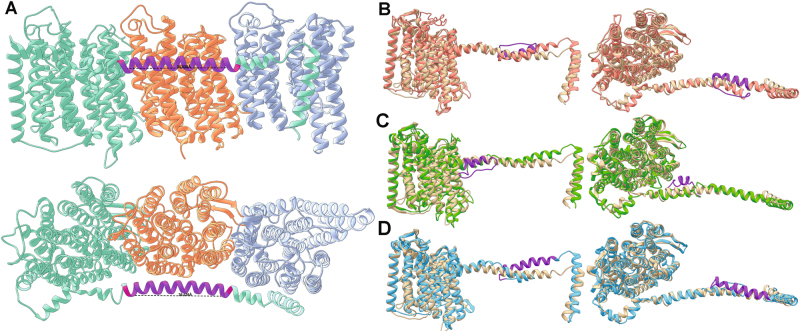
Fig. 6.
Nuo crystal structure and structural homology models highlighting the characteristic amino acid insertion lengthening the amphipathic helical arm that spans the 2M complexes. a NuoL, M, and N subunits from the 4HEA crystal structure with a stretch of 26 amino acids marked in purple on the amphipathic helix (28 denoted by the additional red residues on either end of the purple region). This length of helix is approximately the same width as the NuoM subunit shown in orange. I-TASSER structural homology models of NuoL from Clades 1, 2, and 3 modeled onto the NuoL subunits from 4HEA are shown in b, c, and d, respectively. In all cases the insertions noted in Fig. 5 have been colored purple, and were modeled by I-TASSER as helices that doubled back on the original helix

