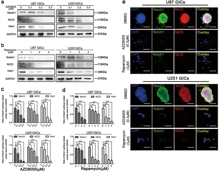
Fig. 5. Autophagy reduced the protein expression of Notch pathway.
a–d U87 and U251 cells were treated the same as in (Picture 2). Protein expressions of Notch1 pathway were detect by western blot. Data are shown as means ± s.d., n = 3, #P = NS, *P < 0.05, **P < 0.01, Student’s t-test. e Immunofluorescence staining of U87 and U251 GICs, which treated by DMSO, AZD8055 (0.3 μM) and rapamycin (3 μM). The nuclei were stained with DAPI and the antibody against Notch1 and NICD. Images were captured by laser confocal microscope ( × 400), scale bar = 100 μm

