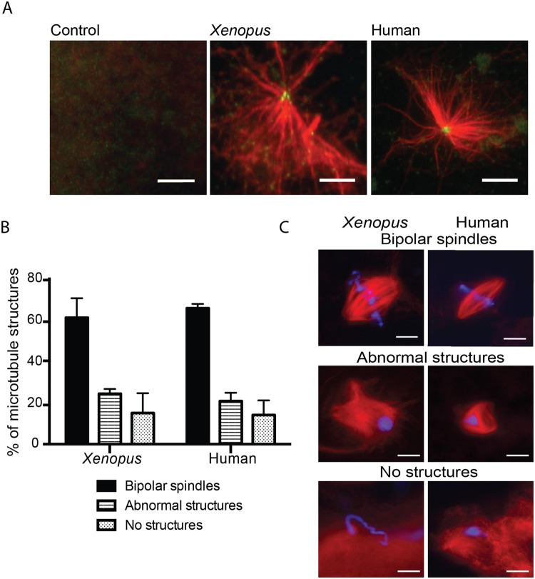
Figure 2.
The human sperm basal body is converted into a fully functional centrosome in the oocyte cytoplasm. (A) Human sperm basal body actively nucleates microtubules in XEE. Immunofluorescence images of KI treated sperm samples incubated with XEE and pure tubulin. Microtubule asters are in red and centrioles in green. Scale bar, 5 µm. (B,C) Capacity of the Xenopus and human spermatozoa to assemble microtubule mitotic structures. The microtubules are in red and the DNA in blue. The structures associated with the DNA were classified as bipolar spindles, abnormal structures and no structures as shown in the images on the right. Scale bar, 10 µm. The graph on the left shows the analysis of 100 and 200 sperm nuclei for Xenopus and human samples respectively. No significant differences were found in any of the samples and microtubule structures. Dispersion data are given as Standard Deviation (SD).

