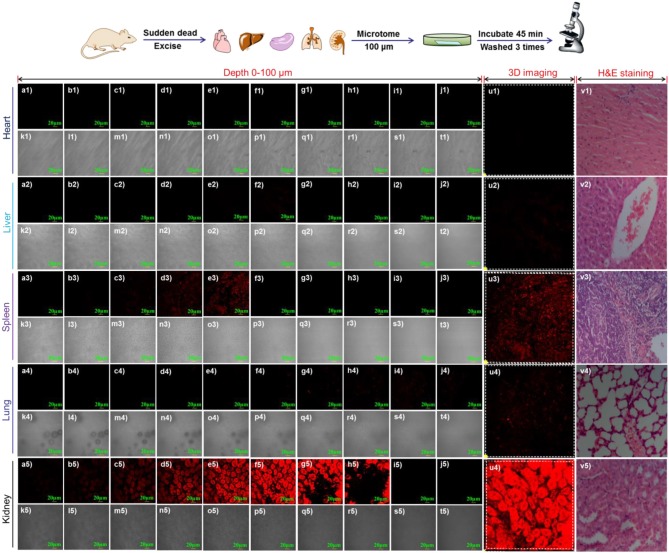
Figure 7.
Confocal fluorescence imaging of endogenous γ-GGT activity in various organs including heart, liver, spleen, lung and kidney. Tissue slices of 100 μm were prepared by freezing microtome (LEICA CM1860 UV). 3D-depth images of different tissue were obtained through z-scan pattern with step size 10 μm. (a-j) fluorescence channel; k-t bright channel; u) 3D-restruction imaging; v) HandE staining. λex = 488 nm and λem = 655-755 nm. Scale bar = 20 μm.

