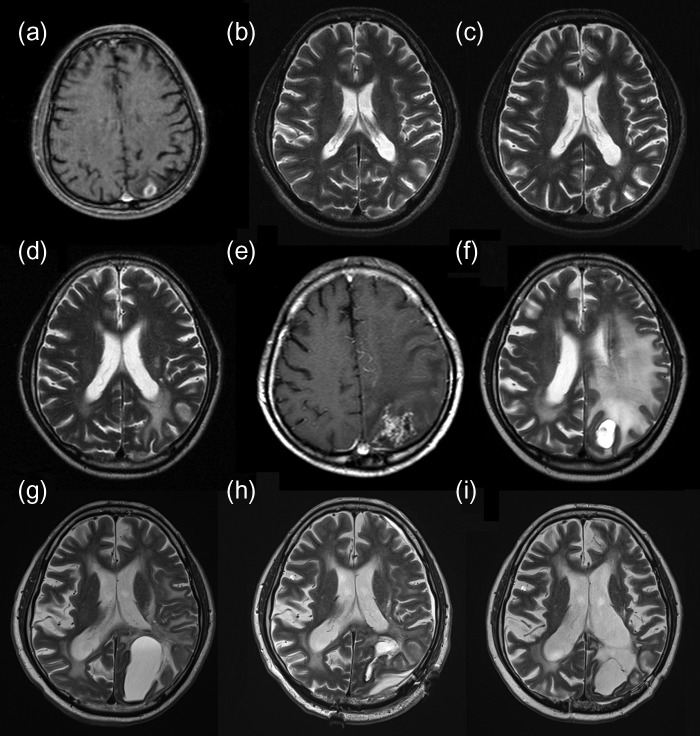
Figure 1:
(a) Enhanced T1WI before SRS revealing a tumor in the left parietal lobe. (b) T2WI (12 months after SRS) without evidence of recurrence or complications. (c) T2WI (35 months after SRS) without evidence of recurrence or complications. (d) T2WI (92 months after SRS) showing a high-intensity area suspected of being a change secondary to irradiation, and no evidence of tumor recurrence. (e) T1WI (118 months after SRS) showing an enhanced lesion with perifocal edema. (f) T2WI (123 months after SRS) showing cyst with an edematous-appearing area. (g) Preoperative T2WI (150 months after SRS) showing expansion of the cyst despite conservative therapy. (h) Postoperative T2WI (151 months after SRS, 1 week after resection) showing cyst shrinkage. (i) Postoperative T2WI (193 months after SRS, 43 months after resection) showing only the removal cavity. T1WI, T1-weighted imaging; SRS, stereotactic radiosurgery; T2WI, T2-weighted imaging.

