Figure 1:

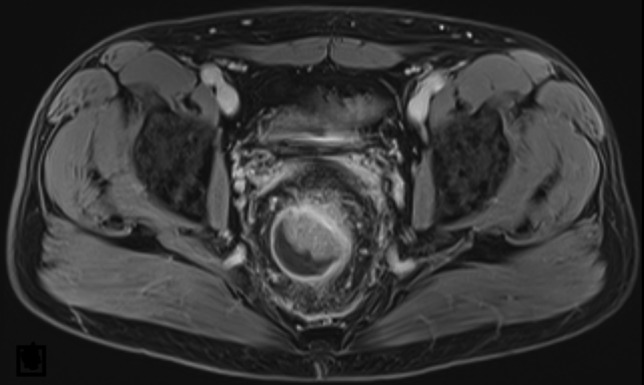
Initial MRI (t2) of the pelvis showing a rectal mass with luminal stenosis and infiltration of the mesorectal fat tissue without involvement of adjacent structures or lymphatic tissue.

Initial MRI (t2) of the pelvis showing a rectal mass with luminal stenosis and infiltration of the mesorectal fat tissue without involvement of adjacent structures or lymphatic tissue.