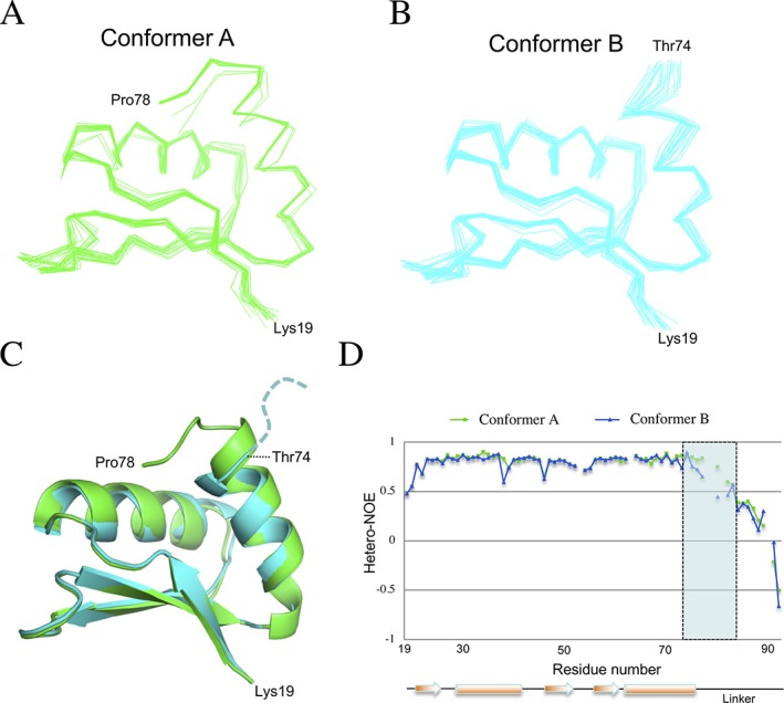
Figure 2.

Structural ensembles of the two PrgK19–92 conformers. Line representation of the backbone atoms for the twenty lowest‐energy models for PrgK19–92 in the (A) conformer A and (B) conformer B, obtained with CS‐Rosetta.20 (C) The two averaged structures are overlaid in cartoon representation, with conformer A in green and conformer B in cyan. The only significant difference lies at their C‐termini, where residues 75–78 of the linker are ordered in conformer A but not in conformer B. (D) The heteronuclear NOE values for the two populations of PrgK19–92 plotted versus sequence, with the secondary structure shown at the bottom. Decreasing NOE values indicate increasing mobility of the 1HN‐15N bond on the sub‐nsec timescale. This confirms that residues 75–78 (shaded rectangle) are ordered in conformer A, but flexible in conformer B
