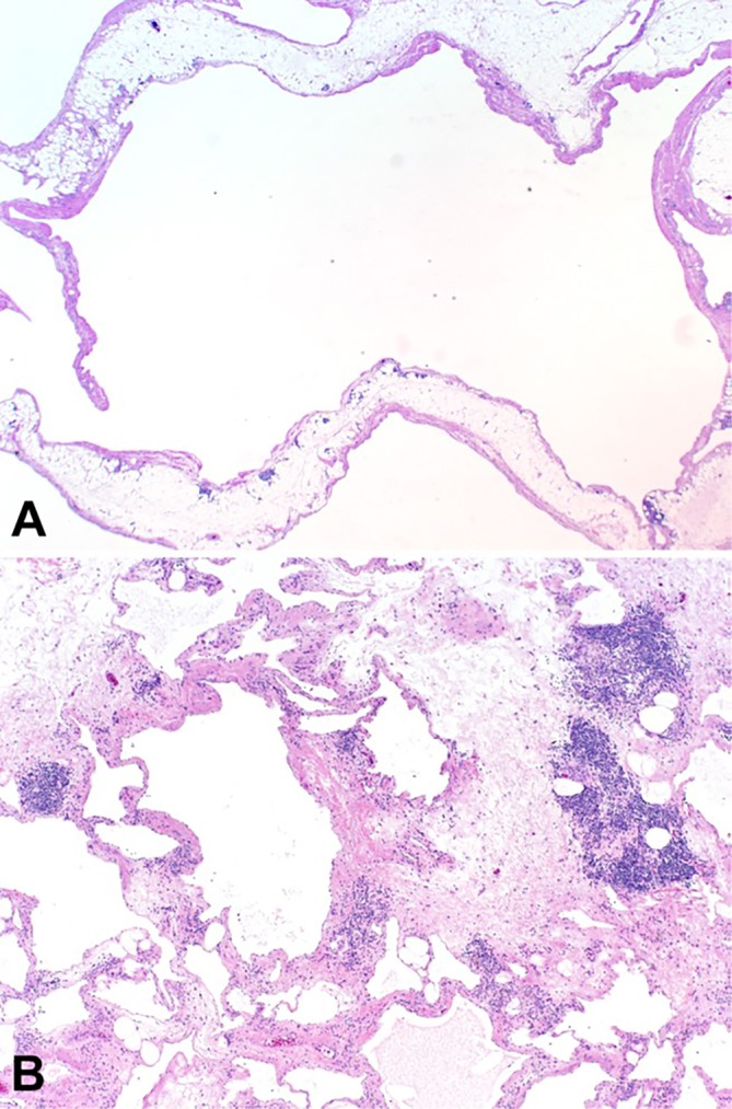
Figure 3.

Routine H&E-stained sections showing (A) large, dilated lymphovascular spaces lined by a simple endothelium, and (B) areas of septal thickening with occasional lymphoid aggregates, interstitial inflammation and focal perivascular smooth muscle (A; 1X, B; 4X).
