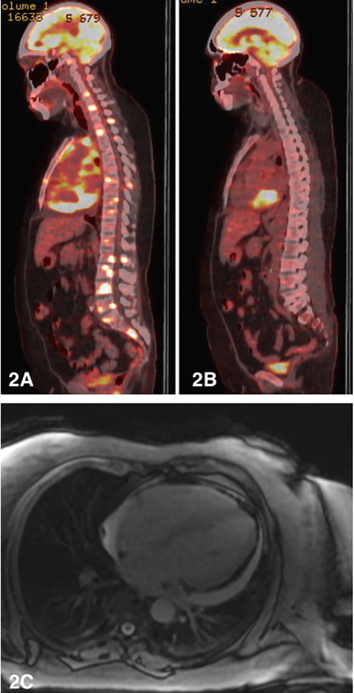
Figure 2.
(A) Initial staging positron emission tomography (PET) scan (sagittal view) showing stage four disease. (B) Postchemotherapy PET scan showing resolution of osseous lesions with persistent high grade metabolic activity in the posteroinferior pericardial recess. (C) Cardiac MRI showing diffuse pericardial thickening and small to moderate circumferential pericardial effusion (12 mm posteriorly).

