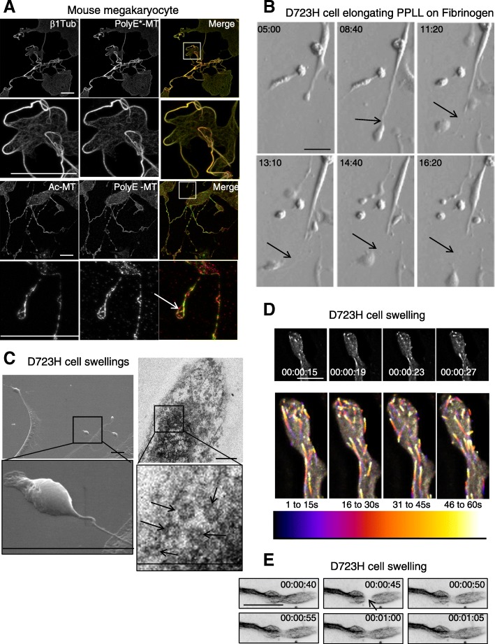
Fig. 1.
Proplatelet MTs are acetylated and polyglutamylated. D723H cells on fibrinogen induce PPLLs containing dynamic MTs. a Maximum intensity projections (MIP) of mature mouse megakaryocytes extending proplatelets stained with β1tubulin or modified MT antibodies. Ac-MT and PolyE-MT patterns are not identical. Low and high magnifications (insets) are shown. Bars 25 μm (b). Modulation contrast time course in hours of cytoplast cleavage at the end of a PPLL extension occurring in D723H cells spread on fibrinogen. Arrows point to the thinning and severing site of the extending protrusion. Bar 30 μm. c Left panels, scanning electron microscopy micrograph of PPLL extending from a D723H cell spread on fibrinogen, showing swellings at their end (top) and detail of a swelling arising from an extremely thin cytoplasmic bridge (bottom). Bars 5 μm. Right panels, transmission electron microscopy micrograph of dense material in a D723H cell swelling (top), higher magnification shows that 5–7 MT bundles are present in the swelling. Bars 500 nm. d, e Fast wide field imaging. d 3D stacks (five slices) derived from live GFP-EB3 comets imaged every second at the tip of a PPLL (MIP). Bar 10 μm. Color temporal projection of MIP indicates comets evolution during temporal windows (1–15, 16–30, 31–45, and 46–60s) and shows MTs enter the forming swellings and coiling back in the shaft of the PPLL. e Live 2D widefield imaging of Sir tubulin-labeled MTs. MT dynamics are observed after FRAPping (for 5 s) the MTs in the PPLL shaft. Bar 20 μm

