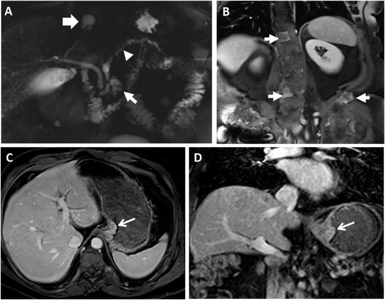
Figure 2.
Representative radiographic GI findings in MAS. (A) MRCP imaging from a 55-year-old man shows diffuse dilation of the main pancreatic duct (arrowhead) along with a cystic lesion in the pancreatic head (thin arrow). A biliary cyst is also seen (thick white arrow). (B) Coronal postcontrast image demonstrates heterogeneously enhancing lesions in the spine and left iliac wing (arrows) in the same patient, consistent with fibrous dysplasia of the bone. (C and D) MRCP images in the same patient who underwent distal pancreatectomy and removal of a gastroesophageal junction polyp are shown. (C) Axial and (D) coronal postcontrast T1-weighted images show the gastroesophageal junction polyp (arrows).

