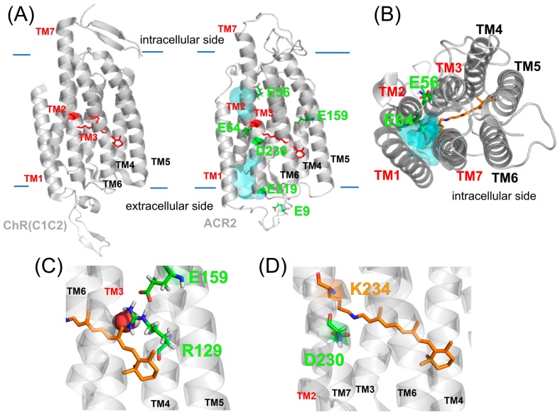
Figure 4.
Homology model structure of ACR2. (A) Crystal structure of the chimeric ChR (PDB; 3UG9) and the homology model of ACR2. In both models, the cavity (cyan colored) seems to be formed by TM1, TM2, TM3 and TM7. (B) Intracellular side of the homology model of ACR2. The cavity is colored cyan. (C, D) Detailed structures around E159 (C) and D230 (D).

