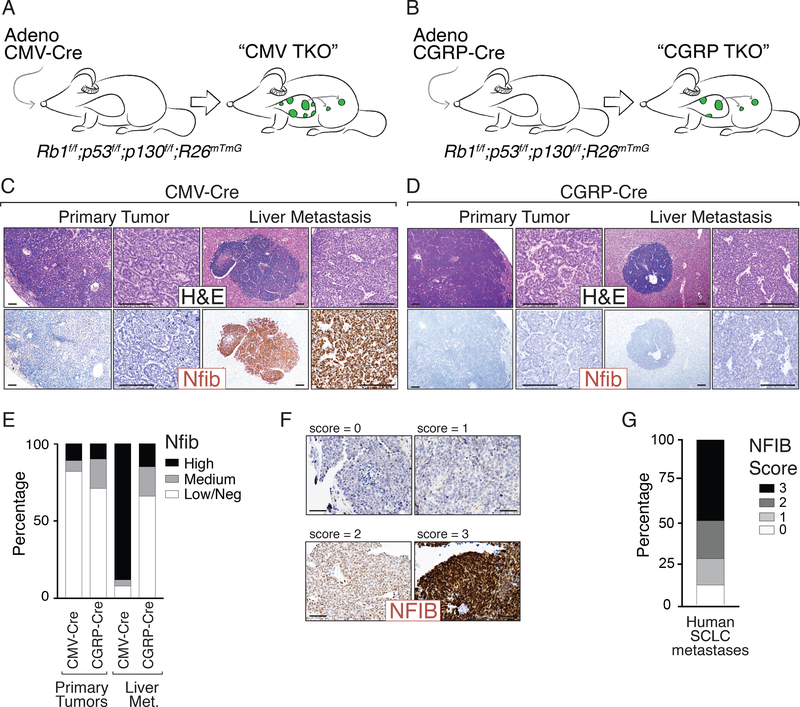
Figure 1: SCLC initiated from pulmonary neuroendocrine cells metastasizes without upregulating Nfib.
A-B. Mouse models of SCLC. Rb1flox/flox;p53 flox/flox;p130 flox/flox;R26mTmG (TKO;mTmG) mice were transduced with either Adeno-CMV-Cre (“CMV TKO”) or Adeno-CGRP-Cre (“CGRP TKO”) to initiate SCLC.
C-D. Representative H&E images of SCLC tumors from CMV TKO and CGRP TKO mice. Immunostaining for Nfib on primary tumors and metastases is shown. Scale bars = 100μm.
E. Quantification of Nfib expression in primary tumors and liver metastases from CMV TKO and CGRP TKO mice. Most of the metastases in CMV TKO mice are Nfibpositive, while most of the in CGRP TKO mice are Nfiblow/negative.
F. Representative immunostaining for NFIB on human SCLC brain metastases. Expression score for NFIB is indicated. Scale bars = 100μm.
G. Quantification of NFIB expression in human SCLC metastases (including lymph node and brain metastases, N=43).

