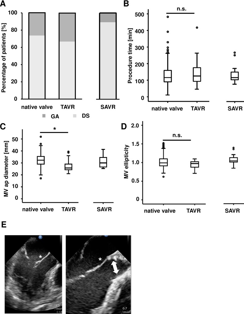
Fig 1. Procedural and echocardiographic characteristics before PMVR.
The patient collective undergoing percutaneous edge-to-edge mitral valve repair (PMVR) was stratified into three cohorts according to a native aortic valve or a previous TAVR or SAVR procedure. A) Percentage of patients undergoing the PMVR procedure in deep sedation (DS) or in general anesthesia (GA). B) Procedure time for PMVR. Boxplots are depicting the median and the upper and lower quartile. C) The mitral valve anterior-posterior (ap) diameter is significantly smaller in patients with previous TAVR. Boxplots are depicting the median and the upper and lower quartile. D) The mitral valve ellipticity index was calculated at baseline. We observed no significant difference between patients with TAVR and without previous TAVR. Boxplots depict the median and the upper and lower quartile. E) Left panel: LVOT view in TEE demonstrating the anatomic situation in a patient with a native aortic valve (white asterisk indicates anterior mitral valve leaflet). Right panel: LVOT view in TEE demonstrating the anatomic situation in a patient after TAVR. Note the close relation of the distal part of the TAVR prothesis and the anterior mitral valve leaflet (white arrow indicates TAVR prosthesis).

