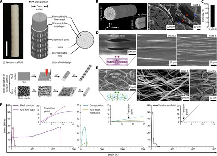
Fig. 1. Fabrication of 3D architecture scaffold with controlled anisotropic microstructure and tendon-like mechanical properties.

(A) Gross appearance of the tendon scaffold designed with core and shell portions building from bioresorbable multilayered PCL fiber meshes and films. Fabrication of the core portion involving steps of uniaxial stretching (s1), layering with a water-soluble poly(ethylene oxide) (PEO) film (s2), and rolling (s3), while the shell portion involving steps of laser microperforation (s1), rolling around a rod to form a hollow tube (s2), and uniaxial stretching (s3). Scale bar, 1 cm. (B) Computed tomography (CT) three-dimensionally (3D) reconstructed and scanning electron microscopy (SEM) cross-sectional images of the tendon scaffold that shows a well-assembled multilaminar structure, with a single film layer constituting the shell portion and multiple fiber mesh layers (separated by PEO interlayers) constituting the core portion. Red and blue arrows (inset 1) representing the fiber and PEO interlayer, respectively. (C) Calculated porosity of the tendon scaffold after dissolving PEO interlayers. (D) SEM images of the shell portion that shows elongated through-holes and orientated ridges/grooves in bulk regions (insets 2 and 3) toward stretching direction (double-headed arrow). (E) SEM images of the core portion that shows orientated waveform fibers (inset 4) toward stretching direction (double-headed arrow) and relatively thicker fibers (inset 5; inactive dashed line) across stretching direction. (F) Representative stress-strain curves of the shell portion, core portion, and tendon scaffold showing reinforced material fabrication. Raw film tube and fiber mesh roll representing control groups of the scaffold shell and core portions, respectively.
