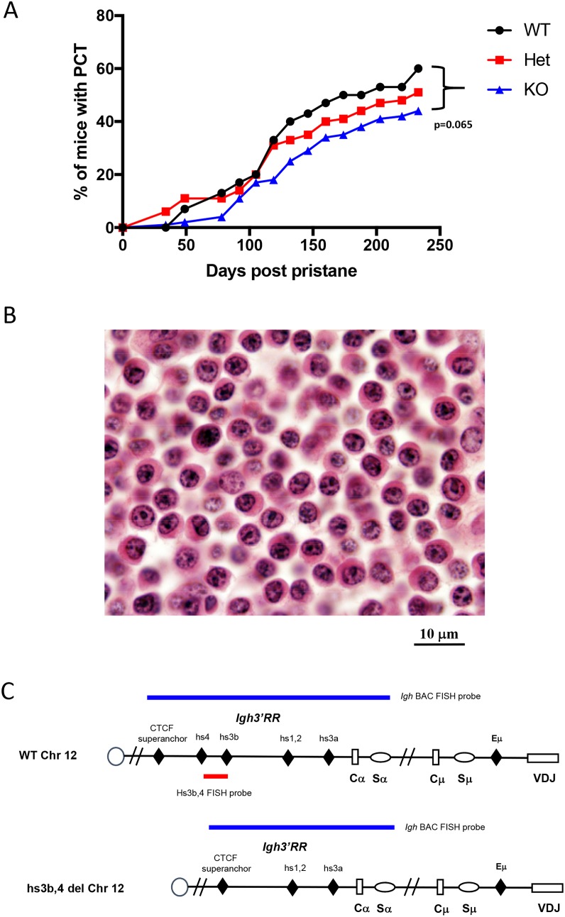
Figure 1. Deletion of hs3b-4 only moderately reduces PCT tumor incidence.
(A) Incidence of PCT in hs3b-4+/+, hs3b-4+/- and hs3b-4-/- mice. The graph shows a lower tumor incidence in the hs3b-4-/- group and a haploinsufficiency phenotype in the hs3b-4+/- group. (B) Photomicrograph of oil granuloma showing advanced growth of a hs3b-4-/- PCT. The tumor has plasmablastic/plasmacytic appearance with abundant eosinophilic cytoplasm and a PCT-typical clock-face nuclear pattern (H&E, 100x oil). (C) Schematics of the WT and hs3b-4-deficient Igh locus. Colored horizontal bars depict location of FISH probes used to detect Igh translocations. 3’Igh BAC probe detects both WT and deleted alleles. hs3b-4 PCR-amplified probe detects only WT allele.

