
Figure 1.
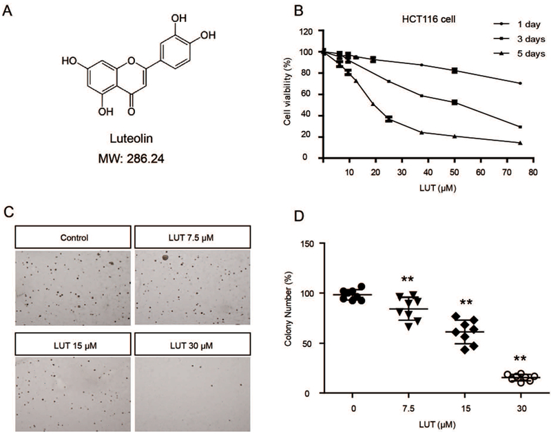
LUT inhibited the cell viability and anchorage-independent growth of HCT116 cells. (A) Structure and molecular weight of LUT. (B) LUT inhibited HCT116 cell viability in a dose-dependent manner. HCT116 cells were plated in 96-well plates at an initial density of 3000 cells/well then treated with various concentrations of LUT for 1, 3, or 5 days. Cell viability was detected using the MTS assay. (C, D) LUT inhibited the anchorage-independent growth of HCT116 cells. Eight thousand cells were seeded in soft agar containing 0.1% DMSO or various concentrations of LUT in 6-well plates for 14 days. Representative images of colonies are shown in C. The relative colony numbers from three independent experiments are shown in D. The data are presented as the mean ± SD. * P < 0.05 and ** P < 0.01 versus the control group.