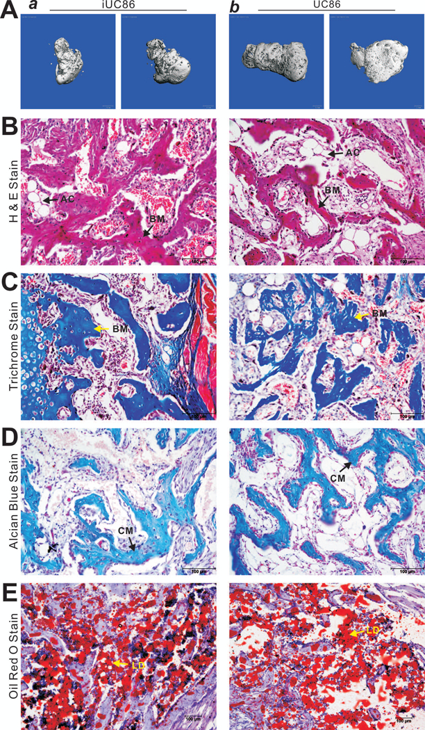
FIGURE 6. BMP9 effectively induces osteogenic, chondrogenic and adipogenic differentiation of the iUC-MSCs in vivo.

Subconfluent iUC86 and primary UC86 cells were infected with Ad-BMP9 or Ad-GFP for 24h, collected and injected subcutaneously into the flanks of athymic nude mice. The animals were sacrificed at 4 weeks after injection. Bony masses at the injection sites were retrieved, fixed and subjected to microCT imaging. Representative images are shown (A). No masses were retrievable from the sites injected with the cells infected with Ad-GFP. After microCT imaging, tissues were decalcified, and either paraffin-embedded for H & E staining (B), Trichrome staining (C), and Alcian Blue staining (D), or frozen-sectioned for Oil Red O staining (E). Representative images are shown. BM, osteoid matrix; CM, chondroid matrix; AC, adipocyte; LD, lipid droplet.
