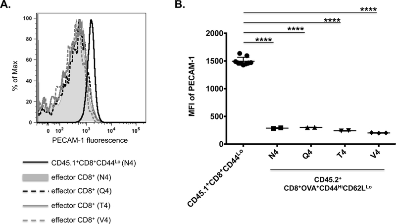
Figure 4. The extent of PECAM-1 down-regulation on activated effector CD8+ T cells in vivo is not affected by the affinity of the TCR for its ligand.

PECAM-1 expression in adoptively transferred CD45.2+ OT-I CD8+ T cells was assessed four days after infection of recipient CD45.1+ mice with Listeria monocytogenes expressing ovalbumin (LM-OVA) containing the native H-2Kb-restricted SIINFEKL peptide (N4), which has high affinity for the OT-I TCR, or its progressively less potent variants SIIQFEKL (Q4), SIITFEKL (T4), and SIIVFEKL (V4). (A) Fluorescence intensity of PECAM-1 expression on splenic CD45.2+ effector (CD44HiCD62LLo) CD8+ T cells from infected mice in one representative experiment; splenic CD45.1+ naïve (CD44Lo) CD8+ T cells from a mouse infected with the N4 variant of LM-OVA served as a control. (B) Statistical analysis of mean fluorescence intensity (MFI) of PECAM-1 expression on splenic CD45.2+ effector (CD44HiCD62LLo) CD8+ T cells from infected mice (n=2 for each variant) relative to CD45.1+ naïve (CD44Lo) CD8+ T cells from the same mice (n=8). Symbols represent individual data points and lines represent means ± SD. Differences between means were determined by 1-way ANOVA and Tukey’s multiple comparison test; ****p < 0.0001.
