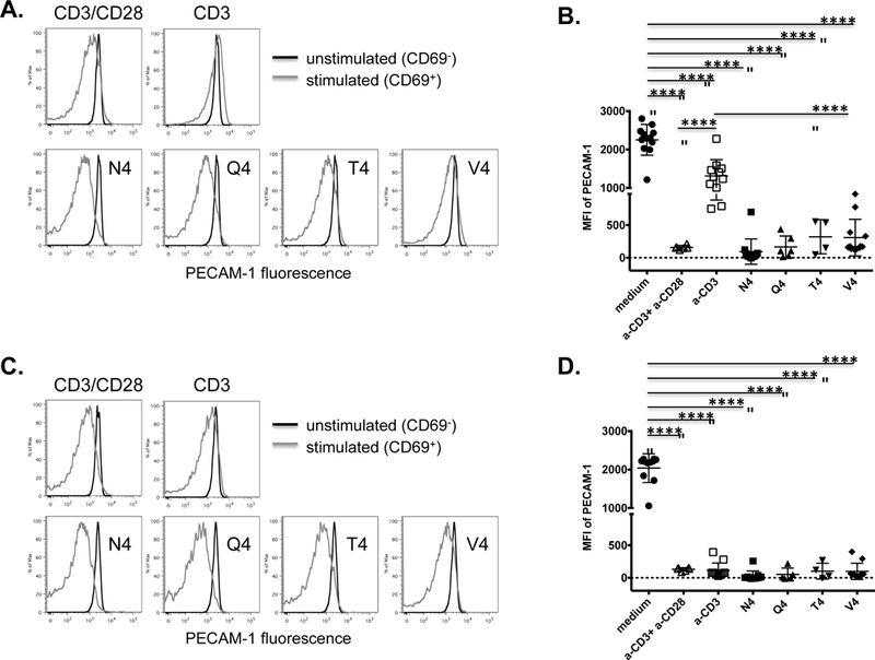
Figure 5. PECAM-1 down-regulation on activated effector CD8+ T cells in vitro requires co-stimulation and is weakly affected by TCR ligand affinity.

PECAM-1 expression on naïve OT-I CD8+ T cells from wild-type (A and B) or DGKζ−/− (C and D) mice was assessed 24 hours after in vitro stimulation, in the presence of antigen presenting cells, with anti-CD3 plus anti-CD28 (αCD3+αCD28), anti-CD3 alone (αCD3), or SIINFEKL peptide variants with progressively lower affinities for the OT-I TCR including native peptide (N4), SIIQFEKL (Q4), SIITFEKL (T4), and SIIVFEKL (V4). Cells cultured in medium alone served as a negative control. (A and C) Fluorescence intensity of PECAM-1 expression on unactivated (CD69-) and activated (CD69+) cells from a wild-type (A) or DGKζ−/− (C) mouse in one representative experiment. (B and D) Statistical analysis of mean fluorescence intensity (MFI) of PECAM-1 expression on stimulated cells from wild-type (B) or DGKζ−/− (D) mice relative to unstimulated cells cultured in medium alone (n=4–11 per condition). Symbols represent individual data points and lines represent means ± SD from at least four independent experiments. Differences between means were determined by 1-way ANOVA and Tukey’s multiple comparison test; ****p < 0.0001.
