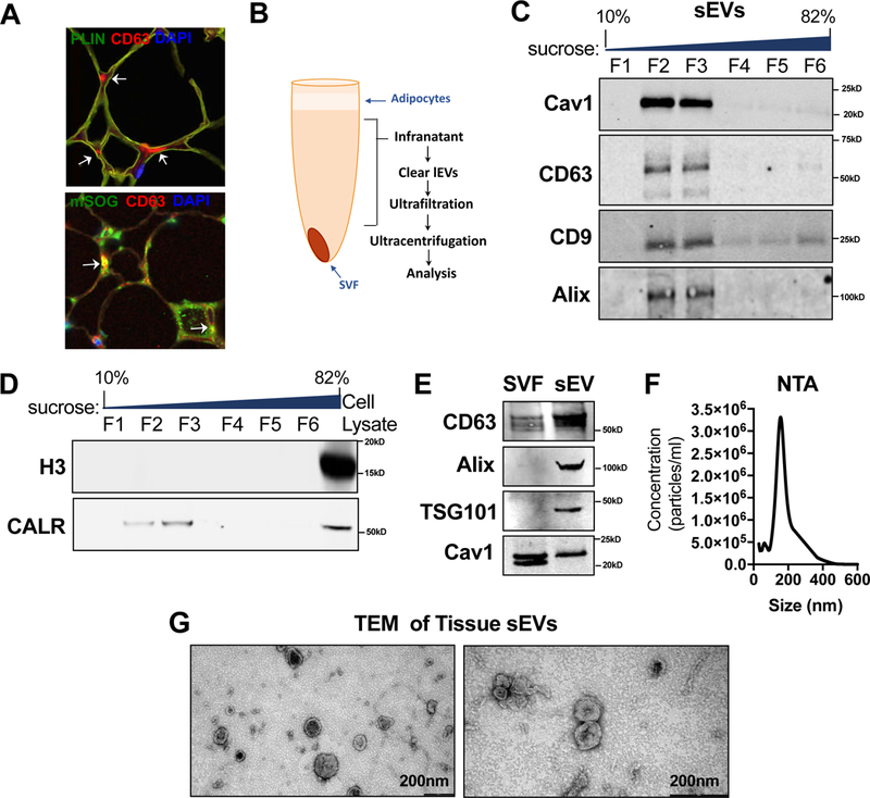
Figure 4: Validation of Tissue sEV Isolation Protocol.

(A) Immunohistochemical staining for the exosomal marker CD63, the adipocyte maker PLIN and miniSOG. Arrows indicate regions of extracellular CD63 staining (red) or CD63-mSOG colocalization (yellow) by confocal imaging. (B) sWAT tissue sEV isolation schematic. (C–D) Western blot of recovered proteins in a sucrose gradient. (E) Western blot detection of cav1 or exosomal makers: CD63, Alix and TSG101 in sWAT SVF or tissue sEV fractions. (F) Nanoparticle tracking analysis (NTA) of purified sWAT tissue sEVs. The experiment was repeated three times. (G) Transmission electron micrograph (TEM) of purified sWAT tissue sEVs.
