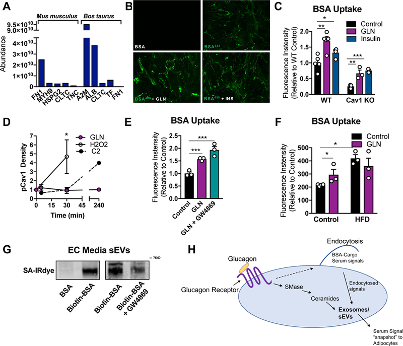
Figure 7: Glucagon Stimulates ECs to Take up Extracellular Components Which are Subsequently Incorporated into sEVs.

(A) mus musculus- and Bos taurus- specific proteins identified by Mass Spectroscopy in AT EC- derived sEVs. (B and C) BSA-conjugated Alexa Flour488 uptake assay was conducted on AT ECs stimulated with either insulin (5 µg/ml) or glucagon (GLN; 10nM). (B) A representative fluorescence image following BSA-Alexa Flour488 uptake. (C) Quantification of Alexa Flour488 intensity (n = 3–4). (D) Western blot densitometry of p-cav1Tyr14 under the indicated conditions. (E and F) BSA-Alexa Flour488 uptake as described in B–C. ECs in E were isolated from mice on a chow or high fat diet (HFD) for 12 wk. (G) ECs were treated with biotinylated-BSA, washed and provided fresh media. Media sEVs were isolated, resuspended in a fixed volume, and analyzed for biotin-BSA by SDS PAGE and streptavidin-IRdye (SA-IRdye) reactivity. (H) Model for the physiological significance of glucagon-stimulated BSA uptake and sEV production. Data is presented as mean ± SEM
