Figure 8.
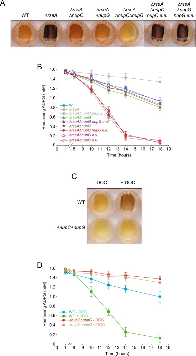
NupC and NupG are the main transport systems involved in the incorporation of extracellular ADPG into envelope stressed E. coli cells. (A) Glycogen iodine staining of ∆rseA, ∆rseA∆nupC, ∆rseA∆nupG and ∆rseA∆nupC∆nupG cells, along with ∆rseA∆nupC cells ectopically expressing nupC and ∆rseA∆nupG cells ectopically expressing nupG (∆rseA∆nupC nupC e.e. and ∆rseA∆nupG nupG e.e., respectively) cultured in solid KM-ADPG. (B) ADPG consumption by BW25113 WT, ∆rseA, ∆rseA∆nupC, ∆rseA∆nupC bearing the empty pSU18 vector, ∆rseA∆nupG, ∆rseA∆nupG bearing the empty pSU18 vector, ∆rseA∆nupC∆nupG, ∆rseA∆nupC nupC e.e., ∆rseA∆nupG nupG e.e., cells cultured in liquid M9-glycerol-ADPG medium. Growth curves are shown in Supplementary Fig. S3G. (C) Glycogen iodine staining of WT and ∆nupC∆nupG cells cultured in solid KM-ADPG with or without 0.1% (w/v) DOC supplementation. (D) ADPG consumption by BW25113 WT and ∆nupC∆nupG cells cultured in liquid M9-glycerol-ADPG medium with or without 0.1% (w/v) DOC supplementation. Growth curves are shown in Supplementary Fig. S3H. In (B,D) values represent means ± SE obtained from four independent experiments with 3 replicates for each experiment.

