Figure 8.
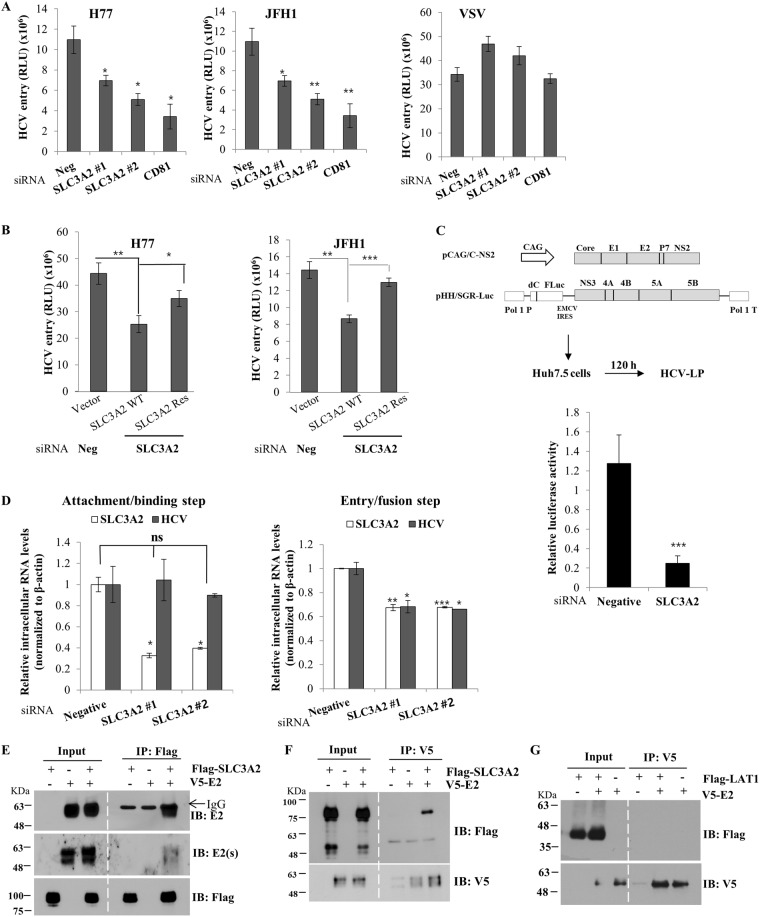
SLC3A2 is required for the entry step of the HCV life cycle. (A) Huh7.5 cells were transfected with the indicated siRNAs. At 48 h after siRNA transfection, cells infected with HCVpp derived from either genotype 1b (H77) or genotype 2a (JFH1) for 6 h. VSVpp was used as a control. Cell medium was replaced with fresh DMEM containing antibiotics. At 48 h postinfection, cells were harvested and then luciferase activity was determined. (B) Huh7.5 cells were transfected with either negative or SLC3A2-specific siRNA for 24 h, followed by transfection with Flag-tagged siRNA-resistant SLC3A2 mutant plasmid. At 24 h after plasmid transfection, cells were infected with HCVpp derived from either H77 (left) or JFH1 (right). At 48 h postinfection, total cells were harvested and luciferase activities were determined. (C) (Left panel) Schematic diagram of the plasmids used for the production of HCV-LP. (Right panel) Huh7.5 cells were transfected with either negative or SLC3A2-specific siRNAs. At 48 h after siRNA transfection, cells were infected with HCV-LP. At 48 h postinfection, total cells were harvested and luciferase activities were determined for single cycle infection. Data represent averages from at least three independent experiments. The asterisks indicate significant differences (***p < 0.001) from the activity for the negative control. (D) (Left panel) Huh7.5 cells were transfected with the indicated siRNAs for 48 h. The cells were incubated with Jc1 at 4 °C for 2 h for binding. The cells were washed with PBS and then bound virions were determined by analyzing HCV RNA levels by qRT-PCR. (Right panel) Huh7.5 cells were transfected with the indicated siRNAs. At 48 h after transfection, cells were incubated with Jc1 at 4 °C for 2 h. The cells were washed with PBS and then temperature was shifted to 37 °C for 4 h. The cells were trypsinized and washed twice with PBS to remove free virions. Internalized HCV virions were indirectly determined by analyzing HCV RNA levels by qRT-PCR. (E) HEK293T cells were cotransfected with Flag-tagged SLC3A2 and either vector or V5-tagged E2 expression plasmid. At 36 h after transfection, total cell lysates were immunoprecipitated with an anti-Flag antibody and bound proteins were analyzed by immunoblot analysis using an anti-E2 antibody and secondary antibodies. To verify E2 protein, we used another kind of secondary antibody, E2(s), which did not recognize heavy chain. (F) Total cell lysates prepared as described in (E) were immunoprecipitated with anti-V5 antibody and bound proteins were detected by immunoblot analysis using an anti-Flag antibody. (G) HEK293T cells were cotransfected with Flag-tagged LAT1 and either vector or V5-tagged E2 plasmid. At 30 h after transfection, total cell lysates were immunoprecipitated with an anti-V5 antibody and bound proteins were analyzed by immunoblot analysis using an anti-Flag antibody.

